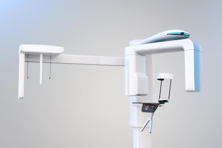
3D Cone Beam
Aspen Dental of Cache Valley offers 3D cone beam x-rays.
This procedure is used when standard x-rays don’t provide dentists with enough information. It shows a more comprehensive image of the patient’s jaw, with a 3-D image of the teeth, nerves, soft tissues, and bone. To prepare for this x-ray, wear comfortable, loose-fitting clothing. You should remove all metal objects such as eyeglasses, piercings, jewelry, hair accessories, removable dental work, and hearing aids. Women need to inform their dentist if they are or could be pregnant. You may need to wear a special gown during the exam, which will be provided by your dentist.
During the procedure, a cone-shaped x-ray beam moves around the patient’s head, taking several pictures, or scans. This allows the dentist to see a three-dimensional image of the patient’s teeth, nerves, soft tissues, and bone. Cone beam x-rays are often used for orthodontic work, like braces, as well as surgical planning, disease diagnosis, and dental implants. Depending on the type of scanner being used, you will either sit or lie down during the procedure. Your dentist will ask you stay very still while the machine rotates around you, so it can produce the most accurate scans. It only takes 20-40 seconds to take a full-mouth x-ray. The exam will not cause any pain or immediate side effects. After the exam, your dentist will analyze the results and then discuss them with you. Because of the focused, cone-shaped x-ray beam, the risks of radiation exposure from this procedure are minimal.


